


Equipment
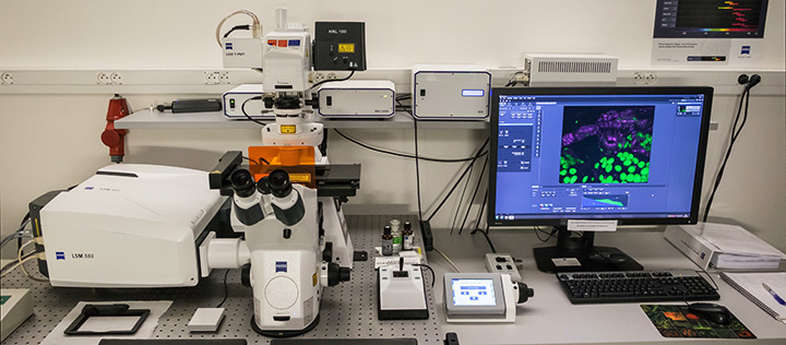
| Inverted laser scanning microscope (2015/2018) | |
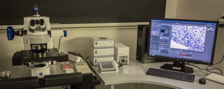
| Description | Zeiss LSM880 laser scanning microscope (Axio Observer Z1, inverted) with Definite Focus 2. System is equipped with high resolution Airyscan detector, spectral GaAsP photodetector, two PMT detectors for confocal, one PMT for transmitted light. All scanners are able to performe bidirectional scanning. Capabilities for FCS, RICS, FRAP, FRET and High Dynamic Range (HDR) applications. |
| Illumination | HXP120 V Illuminator/HAL 100 |
| Confocal lasers: diode laser (405nm-30), Argon ion laser (458, 488, 514nm), DPSS laser (561nm-10), HeNe laser(633nm) | |
| Objectives | Plan-Apochromat 5x/0.16 M27 |
| Plan-Apochromat 10x/0.45 DIC M27 | |
| Plan-Apochromat 20x/0.8 DIC M27 | |
| C-Apochromat 10x/0.45 W DIC M27 | |
| C-Apochromat 40x/1.2 W Kor FCS M27 | |
| Plan-Apochromat 63x/1.40 Oil DIC M27 | |
| alpha Plan-Apochromat 100x/1.46 Oil DIC M27 | |
| Fluorescence filtercubes | FS49/ DAPI (Ex 365 Em BP 445/50) |
| FS38/GFP BP (Ex 470/40 Em BP 525/50) | |
| FS63 HE/mRFP (Ex BP 572/25 Em BP 629/62) | |
| Airyscan emission filters: | |
| BP 420-480 + LP 605 Airyscan | |
| BP 465-505 + LP 525 Airyscan | |
| BP 495-550 + LP 570 Airyscan | |
| BP 555-620 + LP 645 Airyscan | |
| BP 420-480 + BP 495-550 Airyscan | |
| BP 420-4445 + BP 465-505 Airyscan | |
| Signal detection | 1x high resolution Airyscan detector, 1x spectral high sensitive GaAsP photodetector, 2x standard PMT detectors for confocal; one PMT for transmitted light (T-PMT) |
| Additional equipment | Definite Focus 2 - focal drift compensation, active anti-vibration table |
| Software | ZEN black with Experiment Designer, 3D, Tiles, FRAP, FRET, FCS, HDR Analysis modules |
| Location | building B1 room 008 |
| Phone | ext. 830 |
| Reservation | http://rezervace.ueb.cas.cz |
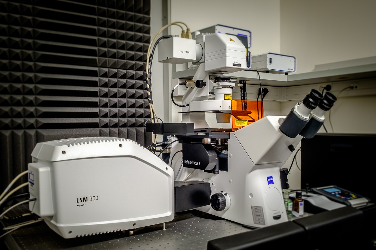
| 2021 | |
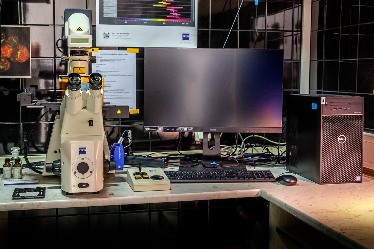
| Description | Zeiss LSM900 laser scanning microscope (Axio Observer 7, inverted) with Airyscan 2 with Multiplex mode. Stabilization in z by Definite Focus.3. System is equipped with high resolution Airyscan2 detector, two GaAsP photodetector, one PMT for transmitted light. Capabilities for FRAP, intuitive FRAP, FRET, photomanipulation, macro module, direct processing. |
| Illumination | Colibri 7 - LED light source (385, 430, 475, 511, 555, 590, 630nm) |
| Laser module LM URGB with diode lasers: 405nm (5mW); 488nm (10mW); 561nm (10mW); 640nm 5mW. | |
| Objectives | Plan-Apochromat 10x/0.45 DIC M27 |
| Plan-Apochromat 20x/0.8 DIC M27 | |
| C-Apochromat 10x/0.45 W DIC M27 | |
| LD LCI Plan-Apo 40x/1,2 Imm Corr DIC M27 | |
| Plan-Apochromat 63x/1.40 Oil DIC M27 | |
| alpha Plan-Apochromat 100x/1.46 Oil DIC M27 | |
| Fluorescence filtercubes | FS49/ DAPI (Ex 365 Em BP 445/50) |
| FS47/CFP (Ex BP 436/20 Em BP 480/40) | |
| FS38/GFP BP (Ex 470/40 Em BP 525/50) | |
| FS63 HE/mRFP (Ex BP 572/25 Em BP 629/62) | |
| FS50/CY5 (Ex BP 640/30 Em BP 690/50) | |
| Signal detection | 1x high resolution Airyscan2 detector with Multiplex mode (=Fast Airyscan), 2x high sensitive GaAsP photodetector; one PMT for transmitted light (T-PMT) |
| Additional equipment | Definite Focus 3 - focal drift compensation, active anti-vibration table |
| Software | ZEN blue with 3D, Tiles&positions, FRAP, Macro, Direct processing modules, APEER connector |
| Location | building B1, Imaging facility - room 011 |
| Phone | ext. 831 |
| Reservation | http://www.ueb.cas.cz/if |
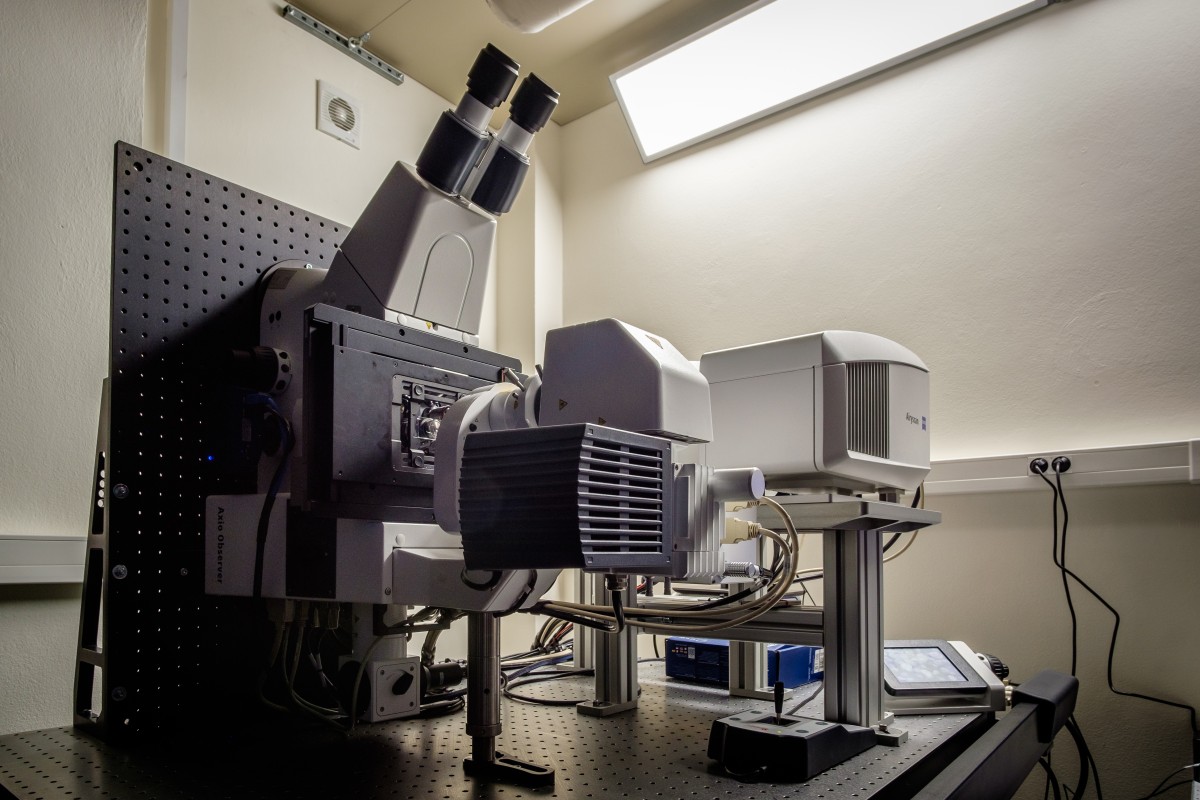
| Inverted laser scanning microscope (2018) | |
| Description | Zeiss LSM880 laser scanning microscope in horizontal setup that enables vertical placement of samples suitable for plants. LSM (Axio Observer Z1, inverted) with Definite Focus 2. System is equipped with high resolution Airyscan detector, GaAsP photodetector, two PMT detectors for confocal, one PMT for transmitted light. Capabilities for 3D, Tiles, FRAP and Deconvolution postprocessing in Zen blue. |
| Illumination | HXP120 V Illuminator/HAL 100 |
| Confocal lasers: diode laser (405nm-30), Argon ion laser (458, 488, 514nm), DPSS laser (561nm-10), HeNe laser(633nm) | |
| Objectives | Plan-Apochromat 5x/0.16 M27 |
| Plan-Apochromat 10x/0.45 DIC M27 | |
| Plan-Apochromat 20x/0.8 DIC M27 | |
| LD LCI Plan-Apo 40x/1,2 Imm Corr DIC M27 | |
| Plan-Apochromat 63x/1.40 Oil DIC M27 | |
| Fluorescence filtercubes | FS49/ DAPI (Ex 365 Em BP 445/50) |
| FS38/GFP BP (Ex 470/40 Em BP 525/50) | |
| FS63 HE/mRFP (Ex BP 572/25 Em BP 629/62) | |
| Airyscan emission filters: | |
| BP 420-480 + LP 605 Airyscan | |
| BP 465-505 + LP 525 Airyscan | |
| BP 495-550 + LP 570 Airyscan | |
| BP 555-620 + LP 645 Airyscan | |
| BP 420-480 + BP 495-550 Airyscan | |
| BP 420-4445 + BP 465-505 Airyscan | |
| Signal detection | 1x high resolution Airyscan detector, 1x high sensitive GaAsP photodetector, 2x standard PMT detectors for confocal; one PMT for transmitted light (T-PMT) |
| Additional equipment | Definite Focus 2 - focal drift compensation, active anti-vibration table |
| CCD Axiocam 506 mono - as alternative for ocular | |
| Software | ZEN black with 3D, Tiles, FRAP Analysis modules. Zen blue with Deconvolution Licence (theoretical and rael PSF, 4 algorithms) |
| Location | building B1 room 009 |
| Phone | ext. 474 |
| Reservation | http://rezervace.ueb.cas.cz |
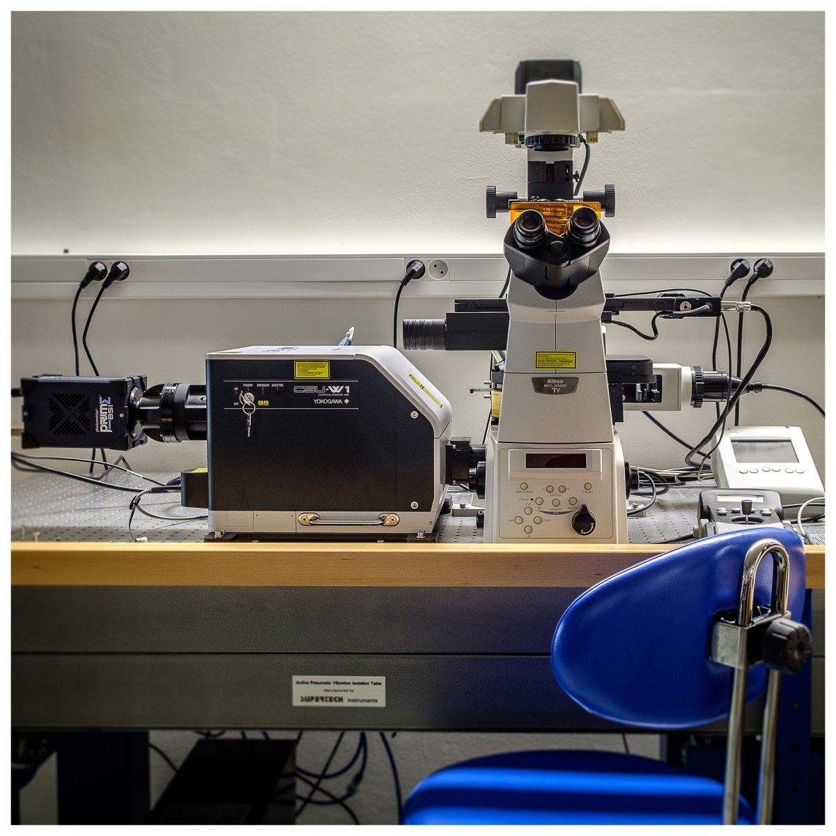
| 2014/upgrade II 2021 | |
| Description | Spinning disc (SD) microscope Nikon (Eclipse Ti-E, inverted) with Yokogawa CSU-W1 SD unit (50mm) in dual camera mode. Highly sensitive backilluminated sCMOS cameras Photometrics Prime BSI are used. Microscope is equipped with perfect focus system, Prior motorized stage with trigered z-Piezo. The setup allows simultaneous two-color imaging with two cameras for pairs GFP/RFP, CFP/YFP and single camera sequential recording of signal in xy, z, lambda, time, positions, tiles and any combination of them. NIS SW 5.30 contains advanced tile scan, FRET measurements, Denoise function, deconvolution, jobs. |
| Illumination | X-cite 120 PC Q Illuminator/HAL 100 |
| Omicron LightHUB ULTRA: 405, 445, 488, 514, 561, 638nm. Laser power: 120mW - 405nm, other 100mW each laser line. | |
| Objectives | Plan-Apochromat L 4x/0.2 |
| Plan-Apochromat L 10x/0.45 | |
| Plan-Apochromat L 20x/0.75 | |
| Plan-Apochromat LS 40x/1.25 WI | |
| Plan-Apochromat L 100x/1.45 Oil | |
| Plan-Apochromat VC 60x/1.2 W | |
| Plan-Apochromat L 60x/1.40 Oil - not mounted | |
| Fluorescence filtercubes | Semrock brightline Em 447/60 (DAPI) |
| Semrock brightline Em 472/30 (CFP) | |
| Semrock brightline Em 525/30 (GFP) | |
| Semrock brightline Em 542/27 (YFP) | |
| Semrock brightline Em 600/52 (RFP) | |
| Semrock brightline Em 641/75 (mCherry) | |
| Semrock brightline Em 708/75 (FarRed) | |
| Signal detection | 2x Photometrics Prime BSI, sCMOS Backilluminated, 2048x2048 pixel, QE 95%, 43/63fps, pixel size 6.5 mm |
| Additional equipment | Perfect Focus System - focal drift compensation; active anti-vibration table |
| Software | NIS Elements 5.30 |
| Location | building B1 room 009 |
| Phone | ext. 474 |
| Reservation | http://rezervace.ueb.cas.cz |
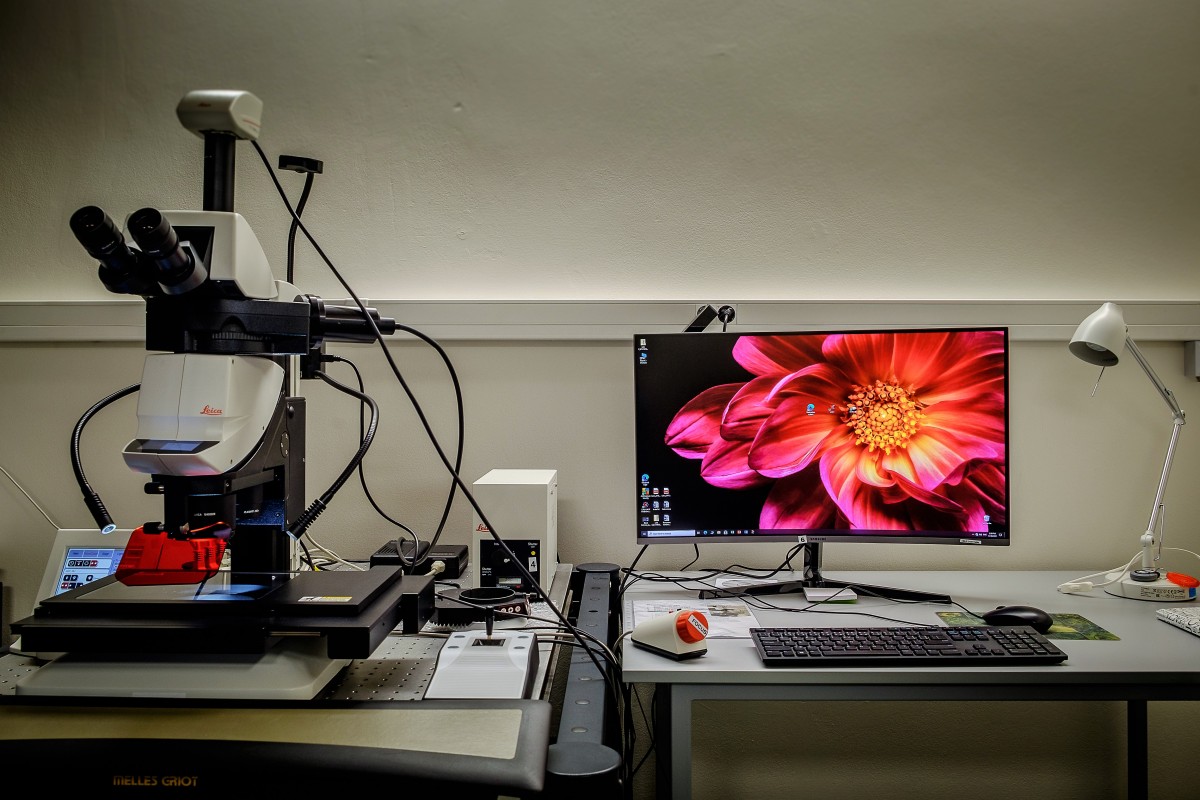
| Upright widefield epifluorescence microscope with optical sectioning using structured illumination (2015) | |
| Description | Upright Zeiss AxioImager Z2 Epifluorescence / Widefield Microscope equipped with ApoTome2 module for optical sectionning using structured illumination. Suitable for general-purpose transmitted light and fluorescence widefield microscopy as well as for the imaging with ApoTome2 structured illumination. Fully motorized. |
| Illumination | HXP120 V Illuminator/VIS LED |
| Objectives | EC Plan-Neofluar 5x/0.16 M27 |
| EC Plan-Neofluar 10x/0.3 M27 | |
| EC Plan-Neofluar 20x/0.50 DIC M27 | |
| C-Apochromat 40x/1.2 W Kor DIC M27 | |
| Plan-Apochromat 63x/1.40 Oil DIC M27 (moved to ApoTome2 microscope) | |
| Plan-Apochromat 100x/1.40 Oil DIC M27 | |
| Fluorescence filtercubes | FS01/DAPI (Ex BP365/12 Em LP 397) |
| FS47/CFP (Ex BP 436/20 Em BP 480/40) | |
| FS09/GFP LP 515nm (Ex BP 450-490 Em LP 515) | |
| FS38/GFP BP (Ex 470/40 Em BP 525/50) | |
| FS43/CY3, Rhodamine, mOrange (Ex BP 545/25 Em BP 605/70) | |
| FS63 HE/mRFP (Ex BP 572/25 Em BP 629/62) | |
| FS25 TRIPLE band/CFP+GFP+mRFP (Ex TBP 400 + 495 + 570 Em TBP 460 + 530 + 625) | |
| Signal detection | CCD Axiocam 506 mono (6Mpix (2752x2208), 19 fps, pixel size 4.5 mm; CMOS Axiocam 105 color (5Mpix) |
| Additional equipment | ApoTome 2; anti-vibration table |
| Software | Zen pro with Deconvolution Module |
| Location | building B1 room 008 |
| Phone | ext. 830 |
| Reservation | http://rezervace.ueb.cas.cz |
| Inverted laser scanning microscope (2006/2020) | |
| Description | Inverted fluorescence widefield microscope Zeiss Axiovert200 equipped with 5Mp colour camera . The setup allows general-purpose transmitted light and fluorescence widefield microscopy in xy, xyz, xyt, xyzt. Suitable for true colour/fluorescence multichannel imaging (e.g. GUS/GFP). |
| Illumination | X-cite series 120 Illuminator/HAL 100 |
| Objectives | EC Plan-Neofluar 5x/0.15 |
| EC Plan-Neofluar 10x/0.30 M27 | |
| Plan-Apochromat 20x/0.8 DIC M27 | |
| C-Apochromat 40x/1.2 W Corr FCS DIC M27 | |
| Plan-Neofluar 40x/0.75 | |
| Plan-Apochromat 63x/1.40 Oil DIC M27 | |
| Fluorescence filtercubes | FS49 DAPI, Hoechst (Ex 365 Em BP 445/50) |
| FS01/DAPI Long Pass (Ex BP365/12 Em LP 397) | |
| FS38/GFP, FITC BP (Ex 470/40 Em BP 525/50) | |
| FS20/Rhodamine, TRITC, mOrange (EX BP 546/12 EM BP 575-640) | |
| Signal detection | CMOS camera Axiocam 705 colour: 2464 x 2056 pixels; pixel size 3.45 mm, binning |
| Additional equipment | Stage holder for multiwell plates. LaCon POC-R-cell cultivation system with peristaltic pump and heating block. |
| Software | Zen Blue |
| Location | building B1, 1st floor, room 112 |
| Phone | ext. 474 |
| Reservation | http://rezervace.ueb.cas.cz |
| 2020 | |
| Description | Fully motorized fluorescence stereomicroscope with motorized table and imaging system. Many illumination and polarization options: transmitted light: bright field to dark field; Rottermann contrast; LED top illumination: ring light / ring light with diffuser; spot light illumination (SLI) ("gooseneck"). The setup allows light and fluorescence microscopy in xy, xyz, xyt, xyzt. Designed for the detection/screening of fluorescence in larger objects with the possibility of microscopy on closed plates (seedlings, callus, seeds, leafs). Moss microscopy. |
| Illumination | HXP120 V Illuminator/HAL 100 |
| Objectives | Plan-Apochromat 1.0x |
| Plan-Apochromat 0.63x | |
| Plan-Apochromat 2.0x | |
| Fluorescence filtercubes | M205FA/165FC: |
| Filter set DAPI/GFP/mCHerry ET403.5/23; 484/22; 552.5/17x ET450/20; 518/27; 595/43 | |
| Filter set ET DAPI Ex350/50x Em460/50 | |
| Filter set ET CFP ET436/20x ET480/40 | |
| Filter set ET GFP Ex470/40x Em525/50 | |
| Filter set ET CY5 ET620/60x ET700/75 | |
| Filterset ET RFP ET546/10x ET605/70 | |
| Filter set ET mOrange blocks mCherry | |
| Filter set ET mCherry ET560/40x ET630/75 | |
| Signal detection | Leica DMC6200 colour camera 2.2Mp |
| Additional equipment | Polarization accessories: polarization filters for objective, for ring light and for transmitted light. |
| Software | LasX Premium with EDF, 3D Visual basic |
| Location | building B1, room 009 |
| Phone | no |
| Reservation | http://rezervace.ueb.cas.cz |
| 2019/2021 | |
| Description | Imaging system capable of detailed imaging of the seedlings growth dynamics, development of apical hook or phototropism in different light lengths. The core system consists of a one-board raspberry Pi 3B+ computer, IR sensitive 8MP Pi NOIR v2 camera module, and 875nm IR LED backlight illumination. WS2812B RGBWW LEDs serve as a source of side illumination for the phototropic experiments. The whole system is highly customizable - any desirable source of light can be added. Compact size and flexible controls allow to use them in the phytotrons or fridges. |
| Illumination | HSDL-4220 IR 875 nm LEDs; WS2812B RGBWW LEDs |
| Signal detection | Raspberry Pi NoIR V2.1 8MP 1080P Camera Module |
| Additional equipment | Raspberry PI3B+ 1GB RAM, Waveshare 4,3" DSI LCD touch display, IPS, 800×480, I2C, MIPI DSI |
| Location | Cultivation room 1 |
| Reservation | http://www.ueb.cas.cz/if |
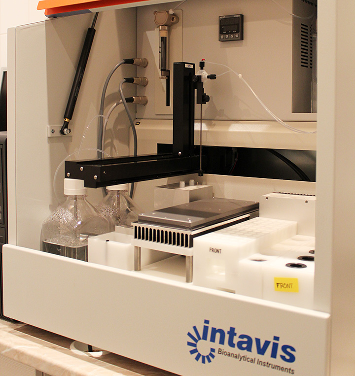
| 2008 | |
| Description | Automated pipetting station for in situ hybridizations and immunohistochemical methods. This machine can be used for stainings of both whole mounts and sections on slides. Size of incubation baskets is suitable for Arabidopsis thaliana seedlings and many other materials. Simultaneous incubation of 60samples (objects or slides) is possible with optional cooling or heating of incubation buffers and incubation area. It is possible to recover washing solutions (e.g. antibodies) and use them repeatedly. |
| Location | building B1, 2nd floor, room 216 |
| Phone | ext 435 |
| Reservation | http://rezervace.ueb.cas.cz |
| 2022/IEB | |
| Description | Windows 11 Pro s instalovaným Windows 10 Pro |
| Procesor: Intel® Core i9-12900K | |
| Paměť: 64 GB DDR5 | |
| Pevný disk: 1 TB M.2 SSD | |
| Optická mechanika: neobsahuje | |
| Grafická karta: NVIDIA RTX A4000/16GB | |
| LAN, WiFi 6E, 1x USB-C, 7x USB 3.2, 3x USB 2.0, 6x DisplayPort, klávesnice, myš | |
| Záruka: 3 roky (následující pracovní den na místě) | |
| Monitor: 2x LCD HP U27 | |
| Location | building B1, Imaging facility - room 011 |
| Phone | ext. 831 |
| Reservation | http://rezervace.ueb.cas.cz |
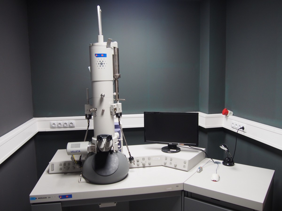
| 2012/OPPK | |
| Description | Up to 100 kV TEM with a tungsten filament electron beam source and a 2k x 2k Side-Mounted TEM CCD Camera. Microscope also has a cold trap for liquid nitrogen, that improves vacuum conditions around the specimen area. The microscope is well suited for routine sample screening and the acquisition of micrographs of good quality with a magnification range from 25 - 200.000 x. |
| Illumination | High Voltage range: 40 to 100 kV in steps of 10 kV, High tension conditioning up to 105 kV, Beam Current: Up to 170 µA, Illumination systém: Two illumination lenses, Condensor 1 and 2 |
| Objectives | |
| Objective Lens: F = 1.6 mm / Cs = 1.6 mm / Cc = 1.5 mm | |
| TEM point resolution: 0.45 nm | |
| TEM line resolution: 0.34 nm | |
| Minimum probe size: 0.50 µm | |
| Smallest focus step: 5.0 nm | |
| Specimen tilt +/- 6° | |
| User-selectable automatic Intensity Limit to prevent electron beam intensity overload on the specimen. User-selectable INTENSITY ZOOM links the illumination control to the image magnification in order to maintain a constant screen brightness during magnification changes. | |
| Signal detection | The Veleta camera - side-mounted tem camera offering a Peltier-cooled CCD chip with 2k x 2k pixels and a dynamic range of 14 bits. |
| Software | Morgagni User Interface |
| Location | building B2, room 016 |
| Phone | ext. 807 |
| Reservation | http://rezervace.ueb.cas.cz |
| 2023/IEB | |
| Modules: Tracking; Exchange objects; Batch Analysis | |
| SW Converter Pro | |
| SW Arivis Virtual Reality | |
| Location | building B1, Imaging facility - room 011 |
| Phone | ext. 831 |
| Reservation | http://rezervace.ueb.cas.cz |
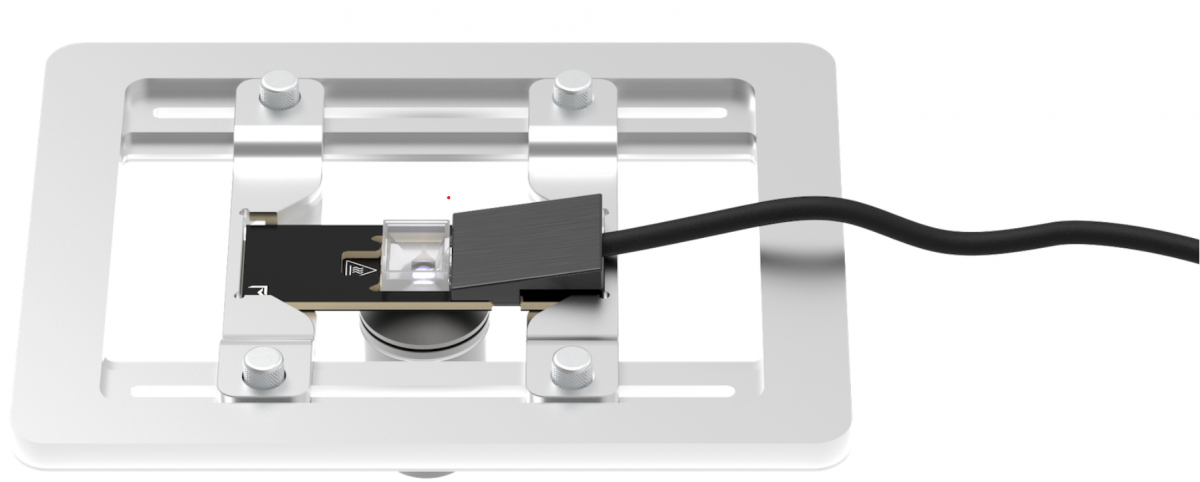 Vaheat - temperature control unit for optical microscopes. Made for investigations of temperature-sensitive processes.
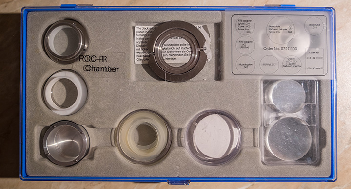 LaCon POC-R-cell cultivation system with peristaltic pump and heating block (chamber)
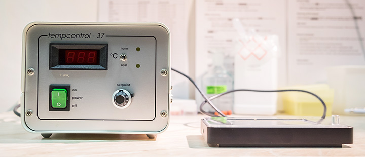 LaCon POC-R-cell cultivation system with peristaltic pump and heating block (heating block)
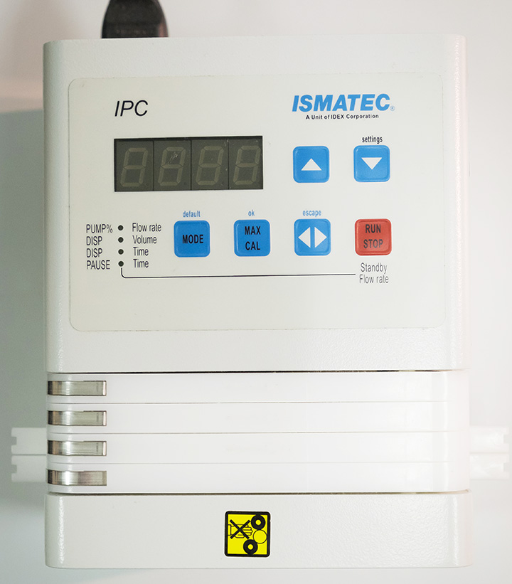 LaCon POC-R-cell cultivation system with peristaltic pump and heating block (pump)
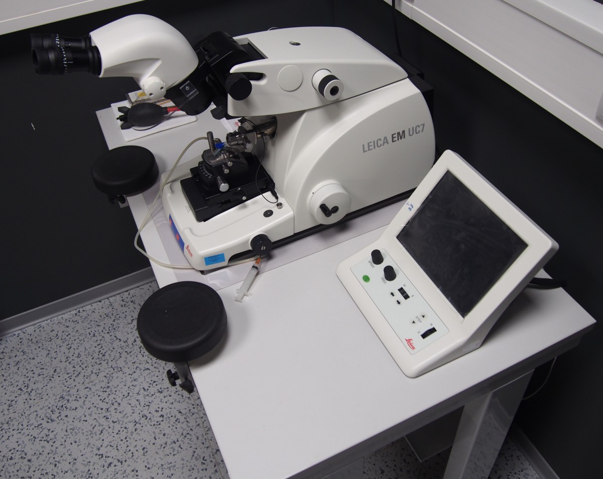 Ultramicrotome Leica UC7